


Cytogeneticists arrange and examine the 23 pairs of chromosomes to zero in on genetic variations. This work can aid in diagnosing cancer, as well as developmental delay, autism, recurrent fetal loss, and other congenital anomalies.
Cytogeneticists, geneticists who study the chromosomes in cells, have a profession with a nifty name and a big job looking at small details.
Within a human cell, cytogeneticists arrange and examine the 23 pairs of chromosomes to zero in on genetic variations. In diagnosing cancer, developmental delay, autism, recurrent fetal loss, and many other congenital anomalies, they do important work that can get to the center of it all. After all, chromosomes, those thread-like structures of protein and nucleic acids in the middle of most cells, hold the genetic keys to a person’s existence.
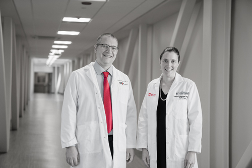
Allen Lamb, PhD, is one of those cytogeneticists and a founding fellow of the American College of Medical Genetics and Genomics, as well as section chief of Cytogenetics and Genomic Microarray at ARUP Laboratories. He specializes in prenatal and postnatal chromosome diagnosis. We talk here with Dr. Lamb about his field, cytogenetics.
- What changes have you seen in the industry?
Genomic microarrays started appearing around 2004–05 and were well-established by 2008–09. They are tools that help us look very carefully for certain changes in the number of copies of a group of genes. These are mutations, but the work is different than DNA sequencing, which looks at specific genes. Cytogeneticists may look at regions of the chromosome that have a few too many genes, for instance. Each microarray is a chip, a small glass slide encased in plastic. The chip’s surface has thousands of synthetic single-strand DNA sequences that form the normal genes in question and can show changes in the number of copies of a chromosome region. These allow us to routinely look for abnormalities unseen by microscopes—gains or losses of chromosomes or parts of chromosomes that are below the resolution of traditional chromosome analysis.
- What is most exciting about this?
With microarrays, we have been able to find new arrangements of genes and characterize new syndromes. For instance, the 22Q (DiGeorge) syndrome, a disorder caused by a deletion on the 22nd chromosome, was better defined by microarray after being found first by the FISH analysis. [FISH is fluorescence in situ hybridization, in which technicians prepare short sequences of single-stranded DNA that match an area of the gene the researcher is looking for—called probes. Fluorescent dye colors are attached to the probes to differentiate them. The probes are then compared with the dual-stranded actual DNA to find differences.] Genomic microarray also helped find 16p11.2, also called Deletion Syndrome, caused by a deletion on a certain part of the 16th chromosome.
- Exactly how much more effective have microarrays been than earlier chromosome analysis?
Before them, a routine chromosome analysis found an abnormality in around 5 percent of cases explored to explain developmental problems. By contrast, microarrays found abnormalities in around 12–15 percent of investigated cases. A lot of discoveries can now happen at the sub-microscopic, sub-visual chromosome level; we previously had very little chance of detecting those. Microarray is quantitative, computer-arrayed analysis. It flags where you should be looking, telling you: Here something is missing, or something extra is over here.
- What is another way genomic microarray pushed previous limits of research?
FISH had helped, but we were only asked to do a FISH analysis by specific request. A clinician needed to look at the overall genotype of the child and order a specific probe, requesting FISH. With microarrays, we are able to study more readily without a specific probe being requested—and we can scan the entire genome.
- What are limits of cytogenetics today?
Microarray has representation of the entire genome, but there are limitations concerning where the individual markers are. There could be something happening between two genetic markers, and it might be difficult to see that area. Microarrays cannot detect single-base changes in the DNA, or balanced rearrangements that may be easily seen by chromosome analysis.
Allen Lamb, PhD"[Because of genomic microarrays], a lot of discoveries can now happen at the sub-microscopic, sub-visual chromosome level; we previously had very little chance of detecting those."
Section Chief of Cytogenetics and Genomic Microarray
- When would you advise testing to proceed beyond cytogenetics to genetic sequencing?
If we get down to the individual gene level and we’re a little uncertain about what is missing in the chromosomes, or about the accuracy of our results, then we might recommend sequencing.
We analyze the significance of a copy number change for the patient. For instance, if the abnormality is a recurrent one, established in constitutional cases regarding child development, we can look to existing literature and make a summary of the apparent cases. We’d do that in order to give the ordering clinician an idea of what to expect in terms of developmental or intellectual abilities. That
said, if the results of our testing are normal, but there is an existing issue in the patient, the clinician may order sequencing.
- What are upcoming changes in the industry?
In genetics over the next four to 10 years, the idea is that we will try to sequence the whole genome or key parts, and in addition to finding small base changes, this should also detect regions that have gains and losses that we traditionally study in cytogenetics. At this point we mainly look at individual base pairs in the DNA by sequencing for finding genetic anomalies causing cancer and constitutional disorders. Eventually, we think molecular genetics and cytogenetics will merge, and more information will result from sequencing. Taking that path is proving a little bit challenging and costly just now. We’re not there yet, but it may happen down the road. As the two fields of molecular genetics and cytogenetics merge, we’ll work to adjust training fellows in those two areas, to blend them together. I think it will take a while before people fully understand both specialties well, but it will happen.
Catherine Arnold, Science Communications Writer