
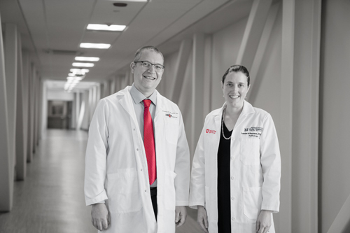


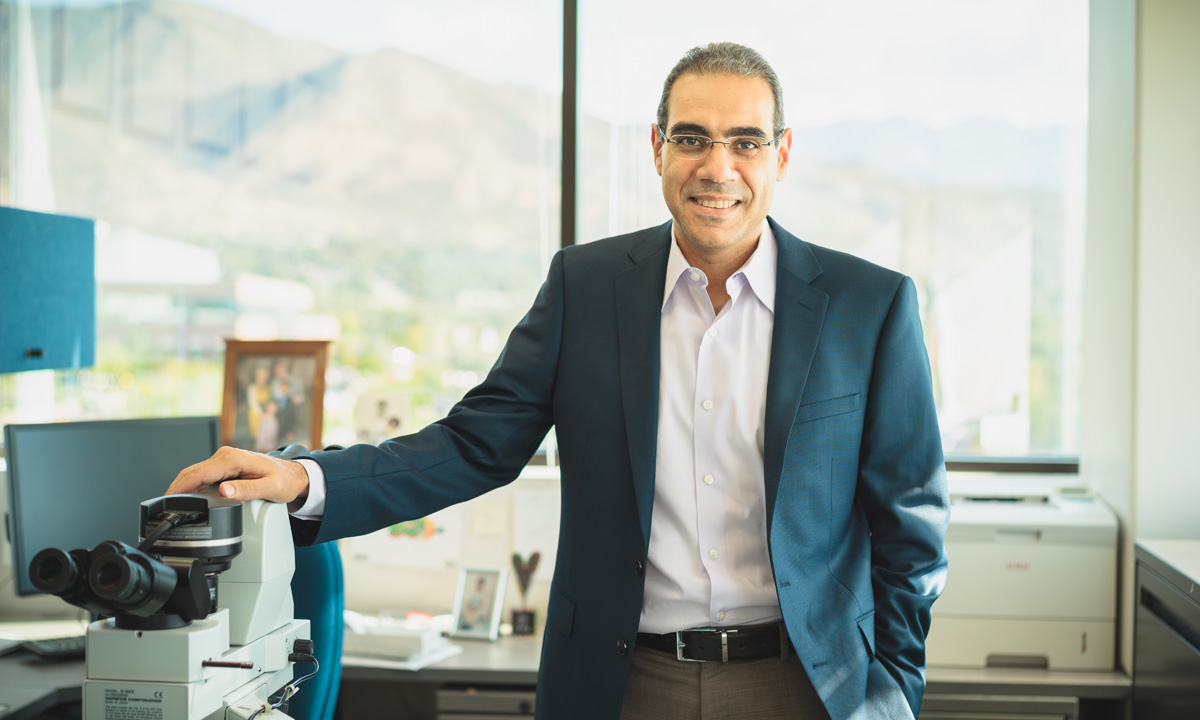
Dr. Salama is a leading force in digital pathology. ARUP’s chief of hematopathology, he is also a professor of pathology and directs the Hematopathology Fellowship Program at the University of Utah School of Medicine.
SALT LAKE CITY, November 21, 2017—ARUP Laboratories and Applied Spectral Imaging (ASI) have teamed up to develop and launch PathFusion™, a cutting-edge solution that combines whole slide imaging, industry-leading tissue FISH analysis, and revolutionary digital tissue-matching of FISH with Brightfield samples. PathFusion provides accurate computer-assisted diagnostics for easy FISH validation, bringing pathology closer than ever to FISH.
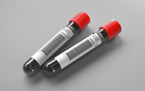
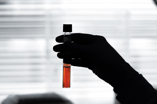
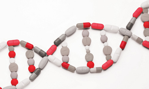
FISH stands for fluorescence in situ hybridization and is a test that maps the genetic material in human cells, including specific genes or portions of genes. Brightfield is the simplest form of optical microscopy illumination techniques (i.e., illuminated from below and observed from above).
Mohamad Salama, MD“We have gone through a vigorous process of validating the system to prove its equivalence to conventional microscopy and have embraced the many benefits it provides, both in clinical results, as well as workflow efficiencies.”
Chief of Hematopathology
Both parties have brought crucial expertise to the table. ARUP is one of the country’s largest academic-medical reference laboratories providing clinical knowledge and workflow experience. ASI is known for its advanced biomedical imaging expertise.
"Our collaboration with ASI represents ARUP’s commitment to embracing the most modern technologies in clinical diagnostics, which we feel make a significant difference for patients,” says Mohamad Salama, MD, chief of Hematopathology at ARUP. “We have gone through a vigorous process of validating the system to prove its equivalence to conventional microscopy and have embraced the many benefits it provides, both in clinical results, as well as workflow efficiencies." Salama is also a professor of pathology at the University of Utah who is often invited to speak at conferences about digital pathology.*
Rapid growth of FISH testing in pathology and the increase in test volumes has created a clear need for advanced digital workflows. PathFusion automates the traditional error-prone process to provide higher diagnostic confidence. H&E slides are scanned, the pathologist logs in remotely and marks the regions of interest onscreen, then the system automatically matches those regions to the same cells on the FISH slide. FISH analysis is performed using proprietary computer-vision algorithms and the final results are presented to the pathologist in a single intuitive view that includes the H&E, IHC, and FISH slides.
"I am excited to be launching PathFusion, which presents a paradigm shift in how FISH pathology is performed," said Limor Shiposh, CEO of ASI. "We joined forces with ARUP to advance this novel tool that, at the end of the day, is critical to patients. ARUP’s deep clinical knowledge and workflow experience, in combination with our imaging and analysis expertise, helped to deliver truly valuable modern innovation."
About ASI: A global leader in the development of biomedical imaging solutions, supporting fluorescent, Brightfield and spectral image-acquisition, for the Pathology and Cytogenetics markets. HiPath Pro™ and PathFusion™, powered by GenASIs™, are automated imaging platforms that provide advanced diagnostic aids for pathologists, with reproducible and standardized results. ASI platforms support manual and automatic scanning for a wide range of workflows and applications, to best suit the needs, size and budget of any laboratory.
ARUP Media Contact:
Peta Owens-Liston, (801) 583-2787, ext. 3635, peta.liston@aruplab.com